Have you ever felt following needs during the studying of the small intestine?
“The results came differently when you are working with cells and animals. Do not know why.”
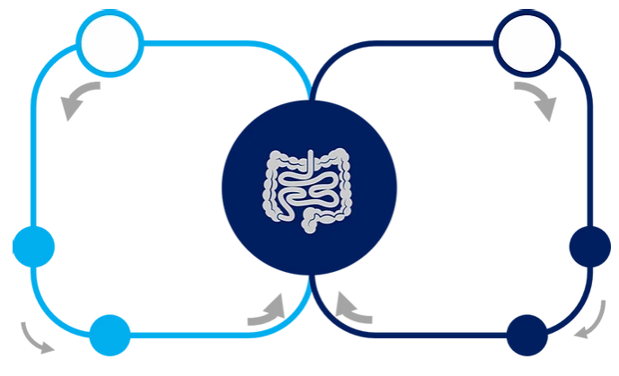
“I want to perform absorption model experiments in the small intestine alone.”
“I want to control drug dosage to the small intestine directly.”
“I want to sample directly from the superior mesenteric vein.”
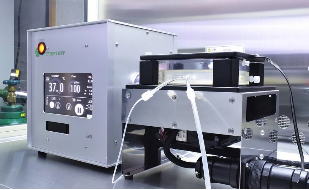
The small intestine chamber of our “Organ Culture System” allows you to run small intestinal perfusion experiments in rats surprisingly easily.
In this issue, I would like to introduce an example of the experiment.
For graphs and tissue slices images can be downloaded here.
Overview
The small intestine was removed from the rat and after 20 hours of perfusion culture in the Organ Culture System, it showed peristalsis and maintained its perfusion rate.
Background and aim
To allow analysis in the isolated small intestine, which is not possible to analyze in individual animals, we aimed to achieve a perfusion culture that remained active and ready for analysis.
The perfusion rate was defined by the following equation.
Perfusion rate = return volume from vein (g)/infused volume (g)
Result
20 hours of perfusion culture, peristalsis was observed, and perfusion while maintaining a perfusion rate of around 40%. It was possible (Fig. 1).
In the perfused tissue sections, smooth muscle and mucosal-like tissue were maintained (Fig. 2).
These results suggest that the tissue section after perfusion maintained its activity and the return fluid from the vein could be analyzed (Fig.2).
The figures are available for download.
References
Sano, K. et al. J Artif Organs (2019) link